 This low spatial resolution is offset to a big degree by improved contrast resolution, which is more pleasing to the attention. Because of their inherent high distinction, direct digital techniques are also becoming the selection imaging device for very giant animals. Higher kV settings produce more penetrating beams in which a better percentage of the x-rays produced penetrate the subject being radiographed. There can be a decrease in the share distinction in absorption between tissue types.
How to Take Perfect Veterinary X-Rays Every Time
Most vets agree that X-rays are secure for pregnant pets once puppies or kittens have gestated for 50 days. Vets X-ray pregnant pets to see how many puppies or kittens the mother is carrying and their positioning. The vet may also compare the fetus sizes to the mother’s birth canal to gauge supply safety. The importance of getting your dog microchipped cannot be exaggerated. Microchips have confirmed time and time once more to get lacking pets residence, typically even years after they go missing. Your dog is unlikely to experience important discomfort during microchipping, and your dog won't ever need their microchip repeated, besides in rare cases. Your vet will totally check your canine for a microchip via the scanner.
 Estos sonidos son normales y se deben al movimiento de la sangre a través de los vasos. El ecógrafo portátil GE LOGIQ y también R8 ofrece diagnóstico en aspecto con la última tecnología en procesamiento de imagen por ultrasonidos que viene de modelos de gama superior. Desarrollado para un empleo pluridisciplinar, es con la capacidad de ofrecer excelentes posibilidades en abdomen y realizar exploraciones cardiológicas de prominente nivel. Se emplea, generalmente, para el estudio de tejidos blandos, como los órganos abdominales, testículos, cuello, ojos, corazón y ciertas construcciones de la cavidad torácica, así como tejidos superficiales (piel y tejido subcutáneo, tejido mamario, etc). Se emplea también para la opinión del sistema musculoesquelético, eminentemente para la exploración de músculos, tendones y ligamentos.
Áreas de aplicación del dispositivo de ultrasonido veterinario.
Se aconseja que todo animal que vaya a ser sometido a una ecografía abdominal, asista a la cita en ayunas (12-18 horas de sólidos, 5 horas de líquidos), tal como con una retención de orina de por favor clique stuart-brennan-2.federatedjournals.com lo menos 5 horas. La línea de base es el eje que representa el cambio de dirección de frecuencia Doppler en un análisis fantasmal y color. Hace aparición con igual espacio para los registros Doppler positivos y negativos. En el momento en que deseamos investigar un trazado con mayor detalle, incrementando el espacio para los registros en evaluación. El Doctor menciona que se tienen que manipular los ajustes especialistas a lo largo del estudio puesto que los pacientes cambian en lo que se refiere a características (peso, altura, etc.). Más allá de que se debe emplear el preset más adecuado, siempre y en todo momento hay que modificar las condiciones del equipo para tener éxito. Igualmente, un pilar fundamental es apoyarse en "una compañía seria, responsable y que nos protega", como Ultravet.
¿Qué es la insuficiencia venosa?
En relación a la glándula adrenal derecha están la arteria frénico abdominal derecha, vaso pocas veces identificable en modo B. El Examen Doppler se utiliza para valorar el estado del sistema circulatorio, advertir anomalías de la salud vasculares, medir el fluído sanguíneo y diagnosticar condiciones como trombosis venosa, insuficiencia venosa y aneurismas. En la región del 12° y 13° espacio intercostal, con la marca del transductor hacia craneal, se observa la vena cava caudal y la arteria aorta abdominal en un corte longitudinal. Se identifica también el lóbulo caudado del hígado y sus vasos parenquimatosos, logrando identificar asimismo en el hilio hepático la llegada de la vena porta y la arteria hepática. En este abordaje exploramos asimismo las arterias celiaca y mesentérica craneal en corte transversal y longitudinal (figura 15). Una ecografía Doppler es un género de ultrasonido que emplea ondas sonoras para enseñar qué tan bien circula la sangre a través de sus vasos sanguíneos.
Iniciemos con una breve introducción a la ecografía
This will improve the number of X-ray photons produced, and thus the overall publicity. We advocate Spot Pet Insurance for these interested in personalized protection. The company’s insurance policies are more customizable than many opponents, with annual restrict choices ranging from $2,500 to unlimited. Spot’s policies additionally cowl a few gadgets that many other pet insurance providers don’t, corresponding to exam fees and microchipping. If your pet starts respiratory abnormally, a chest X-ray can help your vet identify potential well being circumstances like bronchitis, pneumonia or fungal infection. Get fast recommendation, trusted care and the proper pet provides – daily, all year round.
X-Rays for Dogs
However, these procedures often require basic anesthesia or sedation and are extra labor-intensive and costly. Be certain to talk with your veterinarian in regards to the risks and benefits of contrast radiography compared to alternatives such as ultrasound, MRI, and CT. Some veterinary services might base their costs on the dimensions of the dog or the location of the X-ray (e.g., dental vs. abdomen), while others could have a onerous and fast price whatever the view. Even proficient people can miss lesions which might be unfamiliar to them, or so-called "lesions of omission." A lesion of omission is one in which a structure or organ generally depicted on the image is lacking. A good example of that is the absence of 1 kidney or the spleen on an abdominal radiograph. Therefore, explicit consideration to systematic evaluation of the image is very important. It is maybe best to start interpretation of the image in an space that isn't of main concern.
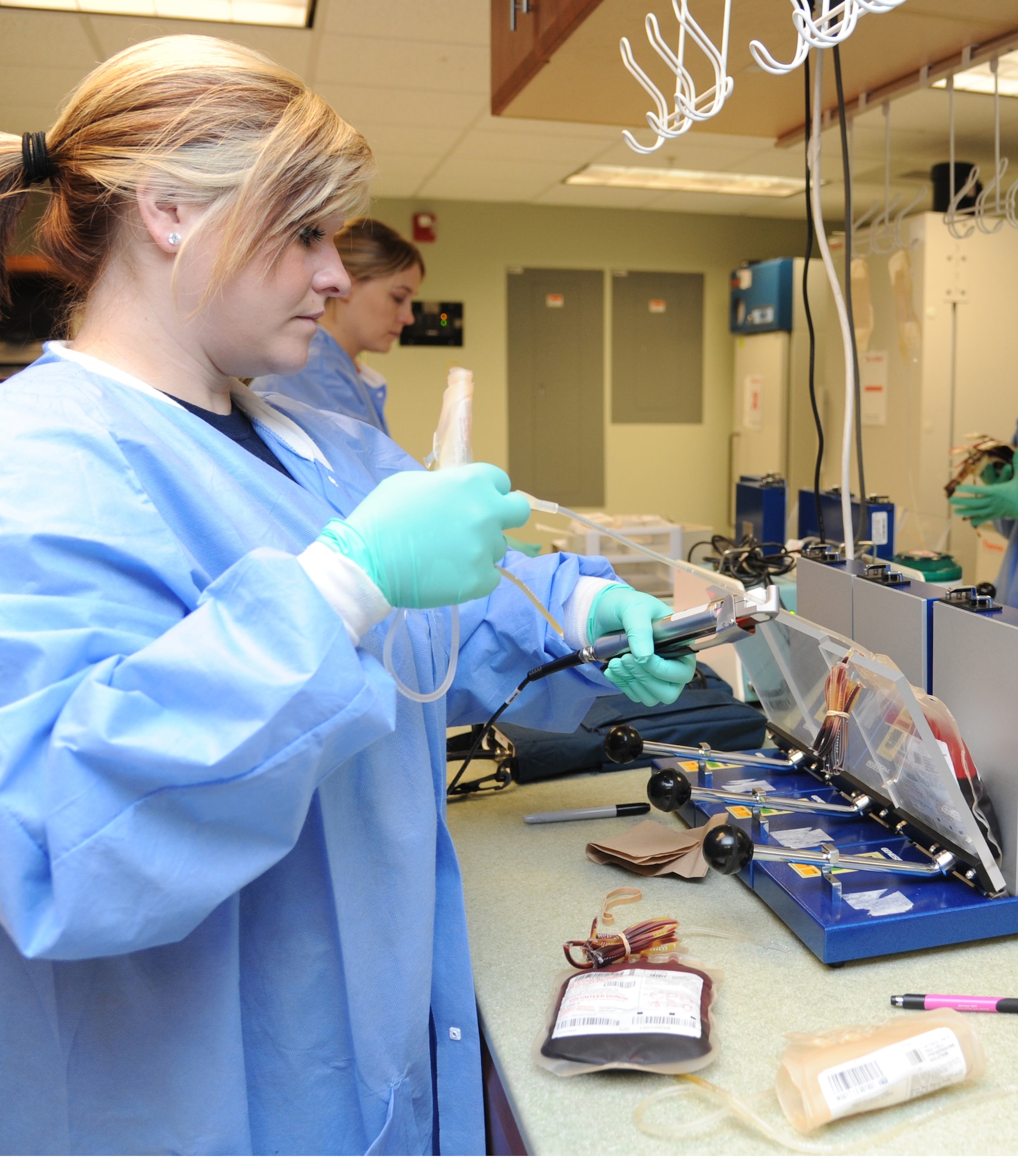
AEC is probably handiest when large numbers of photographs are being carried out of the identical anatomic space by the same personnel. AEC is often not utilized in most veterinary purposes due to the broad variation in physique sizes and conformation of canine. The body’s delicate tissues do not take up x‑rays well and could be difficult to see using this technology alone. Specialized x‑ray methods, known as contrast procedures, are used to help provide more detailed images of physique organs.
If you’re considering giving CBD to your canine, you want to begin by talking to your veterinarian to make certain that your canine is an effective candidate for CBD. The results of CBD are noticeable at variable instances, depending on the product you utilize, the route of administration, the dose required on your dog’s ailment, and the ailment itself. Before purchasing a vet x-ray machine for your clinic, you must take the entire above issues and speak with a educated vendor. An experienced vet x-ray machine vendor will be succesful of think about all your present needs and future progress plans and suggest the greatest possible machine in your specific circumstances. It can also be helpful to record the settings used for each exposure, both on the system or by hand, so with time, we will start to know our machine and what settings work nicely for sure photographs. When thinking about radiation safety, each the patient and the operator, all the time use the lowest potential settings wanted to achieve the diagnostic image.
Radiographic Geometry and Thinking in Three Dimensions
Digital radiographs are saved in a specific format called DICOM (Digital Imaging and Communications in Medicine). This format ensures the security of radiographic photographs taken by stopping tampering with the original picture. The storage of images in DICOM format additionally ensures consistency of file type between all radiographic methods – it is a widespread language’. The uncovered plate is manually placed right into a specialised reader where the inside display screen is scanned by a laser and a digital radiographic image is produced. The last digital picture can then be seen and manipulated utilizing laptop software program (Figure 9). Swallowing a nondigestible object may cause life-threatening well being problems for a pet. An belly X-ray can provide your vet a visible image of the object in your pet’s stomach or intestinal tract to determine whether surgical procedure is important for removing.
Radiography (X-ray)
The larger the kV, the upper their vitality and subsequently their penetrating energy into the patient. Adjusting the kV will enable for changes in each the contrast and exposure of the image produced. Since 1895, when X-rays had been first discovered, radiography has confirmed an invaluable asset in both human and veterinary medication. Digital radiographic images saved in DICOM format are then saved within a PACS community. PACS is the Picture Archiving and Communication System and permits saved photographs to be seen and disseminated to colleagues, referral centres and shoppers. PACS additionally enables the consumer to carry out various functions on the image, similar to zooming, distinction and brightness adjustments, annotations and measurements.
Diagnostic Imaging
In many circumstances, there is good cause to use both X-ray and ultrasound to diagnose or to slender down your pet’s well being concern. For example, if it appears to the vet that the pet ingested a foreign object, then an X-ray would probably be accomplished first. But should that veterinary X-ray present an enlarged spleen, then an ultrasound could be used to get a better picture of the spleen since it is gentle tissue. A main limitation of radiographic imaging is that the pictures are two-dimensional although the affected person is three-dimensional. This signifies that the radiographic look of buildings and/or lesions will rely upon their orientation with respect to the primary x-ray beam and receiver. Consequences of radiographs being two-dimensional are (1) magnification and distortion, (2) picture of a familiar half showing unfamiliar, (3) lack of depth perception, and (4) superimposition. X-rays, put simply, are 2D images of a 3D object, a sort of electromagnetic radiation produced when electrical power from electrons is transformed into X-rays within a specifically designed tube.
X-RAY Systems